Journal of Dairy Science and Biotechnology
Korean Society of Dairy Science and Biotechnology
REVIEW

비활성화 세포, Persister 세포와 VBNC(Viable but Non-Culturable Cells)의 이해

김혜인1https://orcid.org/0009-0008-0893-9916, 송수연1,2,*https://orcid.org/0000-0003-1819-5099
Hyein Kim1https://orcid.org/0009-0008-0893-9916, Sooyeon Song1,2,*https://orcid.org/0000-0003-1819-5099
1전북대학교 동물자원과학과
2전북대학교 농식품융합학과
1Department of Animal Science, Jeonbuk National University, Jeonju, Korea
2Agricultural Convergence Technology, Jeonbuk National University, Jeonju, Korea
*Corresponding author : Sooyeon Song, Department of Animal Science, Jeonbuk National University, Jeonju, Korea, Tel : +82-63-270-2604, Fax : +82-63-270-2612, E-mail : songsy@jbnu.ac.kr

© Copyright 2023, Korean Society of Dairy Science and Biotechnology. This is an Open Access article distributed under the terms of the Creative Commons Attribution Non-Commercial License (http://creativecommons.org/licenses/by-nc/3.0/) which permits unrestricted non-commercial use, distribution, and reproduction in any medium, provided the original work is properly cited.

Received: Oct 04, 2023; Revised: Dec 01, 2023; Accepted: Dec 05, 2023

Published Online: Dec 31, 2023

Abstract

In the field of microbiology, numerous types of bacteria live dormant to survive stresses such as pasteurization and antibiotics. Some bacteria become ‘persisters’ by inactivating their ribosomes, allowing them to ‘sleep’ through stress and revive when the stress has been removed. Under stress, some cells morph into hollow, lifeless structures known as ‘cell shells.’ In microbiology, these cells have been confused with viable cells in the ‘viable but non-culturable cells’ phenomenon. Therefore, this review addressed the concept that when revival occurs, the always-viable persister cells revive, instead of the dead cell husks.

Keywords: stress response; persister cells; viable but non-culturable cells (VBNC); dormant; resuscitation

Persister 세포와 Viable but Non-Culturable Cells 세포

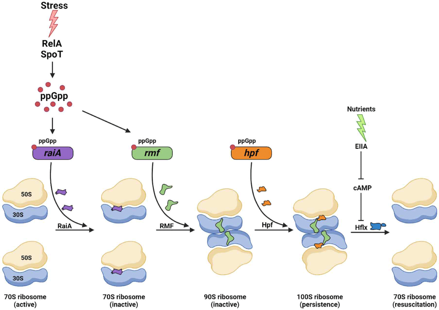
Persister 세포와 VBNC(viable but non-culturable cells), 두 세포의 유형은 대사가 정지되어 있는 상태이나 다시 생존이 가능하다[1]. 이 두 세포 유형의 공통적 특징으로는, persisters[2]와 VBNC[3]가 모두 만성적인 감염에 관여하고, 둘 다 생물막[3,4]에 존재하며, 각각의 스트레스(예, 영양분, 산화성, 산성의 스트레스[1,3,5])에 의해서 생성되며 유제품 공정에 흔히 사용되는 저온살균(pasteurization)에 의해서도 생성된다는 것이 있다. 단일세포 분석을 통해 밝혀진 persister 세포 형성과정에서의 유전적 요소는 Escherichia coli의 RaiA(ribosome-associated inhibitor A), RMF(ribosome modulation fator) 그리고 Hpf(ribosome hibernation promoting factor)와 같은 단백질의 유도로 인한 리보솜 불활성화이다(Fig. 1)[6]. 불활성화된 리보솜은 다양한 스트레스 상황 속에서 alarmone인 (p)ppGpp를 통해 유발되고, (p)ppGpp ribosome dimerization model의 개시를 유도한다(Fig. 1)[7]. 단일세포 분석에서 persister 세포는 휴면기에서 나와 정상적으로 생장하기 위해, 영양소를 발견하고, (p)ppGpp와 cAMP의 농도를 감소시키며[8], 리보솜(ribosome)을 활성화시키는 요소인 HflX(high frequency of lysogeny X)[9]를 통해 리보솜이 다시 활성화된다(Fig. 1)[8]. 이렇게 재생장하는 세포는 주화성(chemotaxis)을 이용하여 영양분 탐색을 시작한다[9]. 반면, VBNC 세포의 경우 형성 메커니즘이 명확하게 밝혀지지 않았지만, 쿼럼 센싱(quorm sensing), 카탈라아제(catalase)와 같은 효소, 항생제, 온도 변화 또는 특정한 탄소원이 VBNC 세포를 재생장하게 한다는 연구가 보고되었다[10]. 하지만 몇 연구에서는 이 연구 결과와 반대되는 연구 결과로 영양분 결핍 스트레스 반응의 주매개자인 (p)ppGpp가 persister 세포에서 증가했을 때, VBNC 세포의 생성이 증가하였으며[11], VBNC는 persister 세포와 다르게 영양분을 공급해도 재생장하지 않기 때문에 VBNC를 휴면기’의 모델로 제시하는 것은 적절하지 않다라는 것이 제기되었다[12]. Wood et al.(2021)의 연구는 VBNC라고 불리는 것들 중 살아있는 부분이 persister 세포이며[1], 배양되지 않는 세포 조각은 죽은 것임을 증명하였다[1]. 20년전, Bogosian et al. (2000)의 연구에서도 마찬가지로 H2O2 스트레스를 제거함으로써 VBNC 세포로 분류된 배양할 수 없는 비브리오균(Vibrio)은 죽은 세포임을 밝혔다[13]. 또한, 콜로니(colony)를 형성할 수 없는 박테리아는 죽었다는 것을 주장하였다[14].

jmsb-41-4-157-g1
Fig. 1. Schematic of the ribosome inactivation and activation cycle in persister cells. Innumerable stress induces RelA/SpoT to produce ppGpp. RaiA, RMF, and Hpf induced by ppGpp each transition the active 70S ribosome to the inactive 70S ribosome, the inactive 70S ribosome to the inactive 90S ribosome, and the inactive 90S ribosome to the inactive 100S ribosome. Persister cells resuscitated when the 100S ribosome into the active 70S ribosome following the decrease in cAMP levels and increase in Hflx production triggered by the removal of stress. RaiA, ribosome-associated inhibitor A; RMF, ribosome modulation fator; Hpf, ribosome hibernation promoting factor; HflX, high frequency of lysogeny X.
Download Original Figure

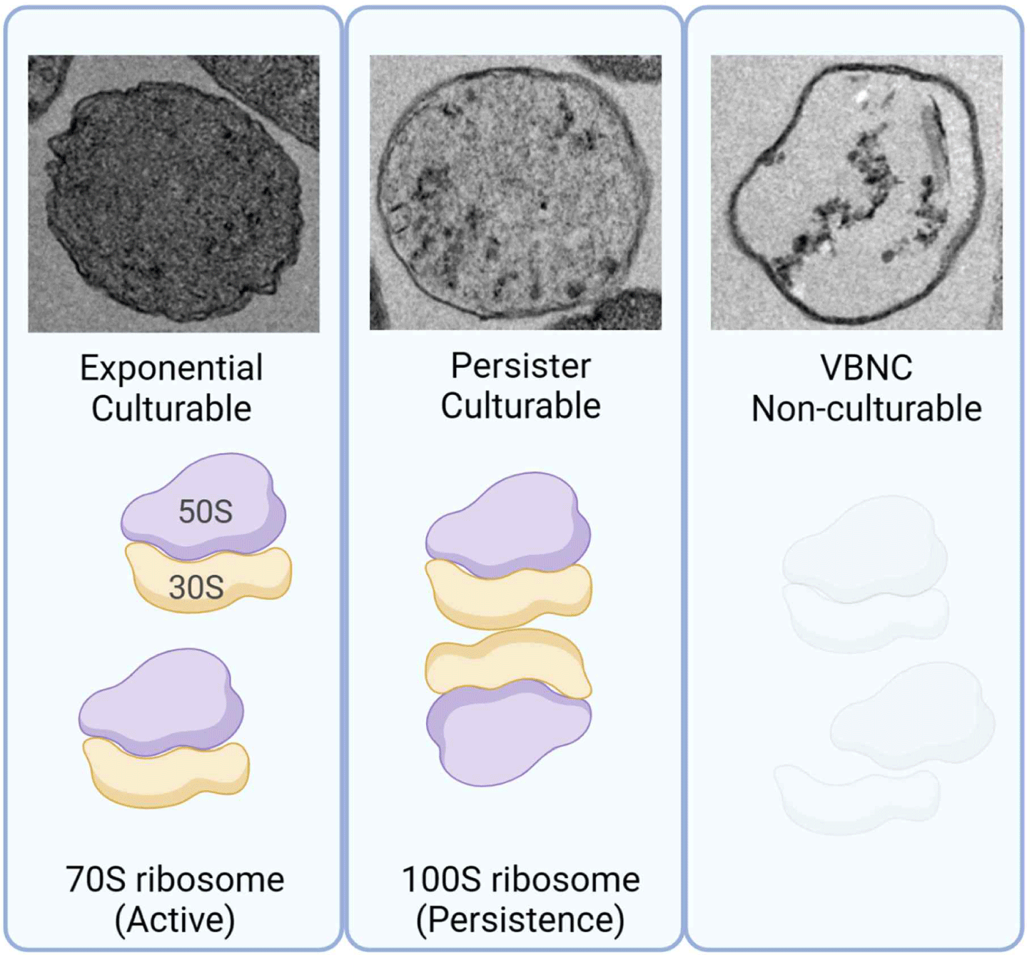
투과전자 현미경을 통한 Viable but Non-Culturable Cells의 필수적 세포물질 결여와 낮은 재활성률

현미경을 이용한 박테리아 세포의 관찰은 VBNC 세포들 중에서 살아있는 persister 세포를 식별하기 위해 중요하다. 투과전자현미경(transmission electron microscope, TEM; Fig. 2)을 통해 E. coli persister 세포는 세포질이 구성성분으로 채워져 있다는 점에서 세포 생장의 지수기에 존재하는 세포와 유사하다는 것을 알 수 있다[1]. 대개 활발히 생장하는 세포일 때는 막대 모양을 가지고 있으나 persister 세포는 회전타원체(spheroid)로 형태를 바꾼다[1]. 이러한 persister 세포는 세포질 구성물인 리보솜을 기반[8]으로 0–6시간 안에 빠르게 휴면기를 벗어나 다시 생장하기 시작한다[1]. 반면, 5주동안의 영양소 결핍으로 형성된 96%의 E. coli VBNC 세포들은 손상되지 않은 세포막을 가지고 있지만, 정상적인 세포질 구성물이 결여되어 있다는 것을 TEM 분석을 통해 관찰할 수 있다(Fig. 2)[1]. 결론적으로 이 ‘세포질 내용물을 가지지 않은 세포 껍질’는 DNA를 포함한 정상적인 세포 요소의 결여로 다시 생장할 수 없다[1]. 게다가, 이 ‘세포 껍질’은 대사 활동이 일어나지 않는다[1]. 두 시간 동안 50°C의 열을 가한 E. coli O157:H7(enterohemorrhagic E. coli, EHEC: 장출혈성 대장균)도 50%가 빈 세포질을 가진 세포 껍질을 형성한다[15]. 그러나 이 중 다시 생장하는 세포는 밀도 높은 세포질을 가진다[15]. 또한, 식물 병원체인 Ralstonia solanacearum은 구리 스트레스로 인해 세포 껍질만을 가진 VBNC 세포가 일부 형성된다[16]. 그러므로 이 VBNC 세포는 다양한 스트레스로 인해 세포질 구성물질이 결여되어 있고, 대사활동이 일어나지 않는, 재생장하지 못하는 ‘세포 껍질’이며 실제로 이러한 특징들은 ‘VBNC 세포가 죽었다.’라는 것을 의미한다.

jmsb-41-4-157-g2
Fig. 2. TEM images of exponential, persisters, and VBNC cells. Exponential cells harvested from grown Escherichia coli K-12 cells at turbidity at 600 nm of 0.8, persister cells induced rifampicin treatment, and VBNC cells induced starvation for 5 weeks in saline solution. Ribosomes indicate each cell's form of ribosomes, either as active 70S particles or hibernated 100S particles. VBNC, viable but non-culturable cells; TEM, transmission electron microscope.
Download Original Figure

세포막 염색으로 구분하는 ‘생존성’의 오류

현재까지 보고된 VBNC에 대한 연구 중 세포막 염색에 대한 해석을 기반으로 VBNC를 구별하는 것은 적절치 않은 것으로 판단된다. 예를 들어 VBNC 세포 관련 연구 중 EHEC(장출혈성 대장균)에 2시간 동안 50°C의 열을 가한 후 VBNC 세포의 수를 알아내는데 PMA(propidium monoazide)-qPCR(quantitative polymerase chain reaction)을 분석 방법으로 사용하였고, 이때 PMA로 염색되지 않은 입자가 VBNC이며 이 세포는 살아있다고 결론 내렸다[15]. 그러나 PMA-qPCR의 결과를 기반으로 VBNC 세포로 판단한 6.5×10⁶ particles mL–1의 세포는 재배양하였음에도 불구하고 다시 생장할 수 없었다[15]. 이 VBNC가 다시 살아날 수 있는 조건은 2시간 미만의 열처리 혹은 더 낮은 열을 가했을 때와 배양 가능한 세포가 존재할 때뿐이다[15]. 즉, 콜로니를 다시 형성할 수 있는 세포는 세포 껍질만을 가진 VBNC 세포가 아닌, 세포질의 밀도가 높은 VBNC 세포만이 생장이 가능한 부분인 것이다[15]. 게다가, 이 연구의 TEM 이미지는 2시간의 열 처리 후, 많은 세포들이 회전타원체가 되고 단백질 응집과 빈 세포질 상태를 보이는데, 이는 확실히 세포가 재생장할 수 없다는 것을 보여준다[15]. 반면에 콜로니를 형성할 수 있는 세포는 손상되지 않은 세포막을 가지고 있고, 이 세포들은 PMA에 의해 염색이 되지 않았으므로 살아있는 것으로 간주되었다[15]. Wood et al.(2021) 연구 그룹은 영양소 결핍으로 형성된 EHEC(장출혈성 대장균) VBNC의 살아있는 부분이 persister 세포라는 것을 규명하였다[1]. 결정적으로, TEM 이미지를 통해서 재생장할 수 없는 세포들은 세포질이 부족하기 때문에 죽은 세포라는 것과, 세포막을 침투할 수 없는 PI(propidium iodide)와 같은 DNA 염색 염료는 세포질의 필수 구성성분의 결여에도 불구하고 손상되지 않은 세포막[1]을 가지고 있으면 DNA가 염색되지 않아 죽었음을 나타낼 수 없기 때문에 VBNC를 연구하는데 적절한 도구가 아니라는 것을 증명하였다.

Viable but Non-Culturable Cells에서 발견되는 응집된 단백질

VBNC의 특징 중 하나로 주장되는 것은 1,000개 이상의 응집된 단백질이 발견되었고 그 응집된 단백질 중에 세포 껍질을 재생장할 수 있게 하는 단백질이 존재한다는 것이다[15,17]. 그러나 변성된 단백질이 세포의 생명을 복원하는 생물학적 과정은 알려지지 않았기 때문에 많은 수의 변성 단백질의 재구성은 불가능한 일이다. 특히 많은 수의 변성된 단백질의 응집은 세포의 죽음을 의미한다[14]. 몇몇 VBNC 세포 관련 연구자들[15,17]과 심지어 persister 세포 관련 연구자들[18,19]에 의한 ‘복원할 수 있는 응집체’와 ‘단백질 응집성’에 대한 주장은 단백질 응집체 데이터[15,17,19]에서 알 수 있듯이 VBNC 세포는 죽은 것임을 확실히 증명한다.

고 찰

국립수의과학검역원(NVRQS)에서 2004년부터 2008년 사이에 435곳의 축사에서 얻은 12,508개의 우유 샘플에서 Enterococci[20]와 Streptococci[21]를 분리해 항생제 내성 실험을 실시하였다. Enterococcus 속에 속하는 105가지 균주 중 약 52%가 7가지 항생제 중 3개 이상의 항생제에 대해 내성을 보였고[20], Streptococcus 속에 속하는 178가지 균주 중 90% 이상이 7가지 항생제 중 최소 1가지에 대해 내성을 보였다[21]. 우유 내 존재하는 두 종의 병원성균 항생제 내성 문제는 유가공 식품의 안전을 위협할 수 있다. 또한, 항생물질의 내성은 2050년까지 1년에 백 만명의 죽음을 예상할 수 있는 매우 심각한 문제이다[22]. 따라서 여러 스트레스와 항생제 스트레스로부터 세포가 살아남는 기작을 명확히 이해하는 것이 필요하다. 이런 이유로 휴면기의 persister 세포와 세포의 빈 껍질(VBNC)을 구별하는 것은 연구의 정확성을 확보하기 위해 중요한 요인이 될 수 있다. VBNC는 감염을 회복시키고 원래의 상태로 돌아갈 수 있는 세포가 휴면 연속체의 일부가 아니며 동시에 오직 persister 세포만이 휴면기 상태이고 다시 생장할 수 있다[1]. 따라서 오랜 기간 동안 휴면 상태로 알려져 온 VBNC는 실제로 죽은 세포이며 VBNC는 persister 세포와 persister 세포가 아닌 세포의 죽음으로 인한 결과이다. 다양한 스트레스에 살아남을 수 있는 휴면기 세포 연구의 명확성을 위해서는 세포가 살아있는지 죽었는지를 결정하는 방법으로 DNA 염색같이 부정확하게 세포 껍질의 손상 여부로 판단하는 간접적인 방법을 활용하기보다는 TEM(Fig. 2)과 생균 계수법을 함께 활용해야 할 것이다.

Conflict of Interest

The authors declare no potential conflict of interest.

References

1.

Kim JS, Chowdhury N, Yamasaki R, Wood TK. Viable but non-culturable and persistence describe the same bacterial stress state. Environ Microbiol. 2018;20:2038-2048.

2.

Mulcahy LR, Burns JL, Lory S, Lewis K. Emergence of Pseudomonas aeruginosa strains producing high levels of persister cells in patients with cystic fibrosis. J Bacteriol. 2010;192:6191-6199.

3.

Li L, Mendis N, Trigui H, Oliver JD, Faucher SP. The importance of the viable but non-culturable state in human bacterial pathogens. Front Microbiol. 2014;5:258.

4.

Spoering AL, Lewis K. Biofilms and planktonic cells of Pseudomonas aeruginosa have similar resistance to killing by antimicrobials. J Bacteriol. 2001;183:6746-6751.

5.

Hong SH, Wang X, O’Connor HF, Benedik MJ, Wood TK. Bacterial persistence increases as environmental fitness decreases. Microbial Biotechnol. 2012;5:509-522.

6.

Song S, Wood TK. ppGpp ribosome dimerization model for bacterial persister formation and resuscitation. Biochem Biophys Res Commun. 2020;523:281-286.

7.

Wood TK, Song S. Forming and waking dormant cells: the ppGpp ribosome dimerization persister model. Biofilm. 2020;2:100018.

8.

Kim JS, Yamasaki R, Song S, Zhang W, Wood TK. Single cell observations show persister cells wake based on ribosome content. Environ Microbiol. 2018;20:2085-2098.

9.

Yamasaki R, Song S, Benedik MJ, Wood TK. Persister cells resuscitate using membrane sensors that activate chemotaxis, lower cAMP levels, and revive ribosomes. iScience. 2020;23:100792.

10.

Dong K, Pan H, Yang D, Rao L, Zhao L, Wang Y, et al. Induction, detection, formation, and resuscitation of viable but non-culturable state microorganisms. Compr Rev Food Sci Food Saf. 2019;19:149-183.

11.

Boaretti M, Lleò MM, Bonato B, Signoretto C, Canepari P. Involvement of rpoS in the survival of Escherichia coli in the viable but non-culturable state. Environ Microbiol. 2003;5:986-996.

12.

Ayrapetyan M, Williams TC, Oliver JD. Bridging the gap between viable but non-culturable and antibiotic persistent bacteria. Trends Microbiol. 2015;23:7-13.

13.

Bogosian G, Aardema ND, Bourneuf EV, Morris PJL, O'Neil JP. Recovery of hydrogen peroxide-sensitive culturable cells of Vibrio vulnificus gives the appearance of resuscitation from a viable but nonculturable state. J Bacteriol. 2000;182:5070-5075.

14.

Baquero F, Levin BR. Proximate and ultimate causes of the bactericidal action of antibiotics. Nat Rev Microbiol. 2021;19:123-132.

15.

Fu Y, Jia Y, Fan J, Yu C, Yu C, Shen C. Induction of Escherichia coli O157:H7 into a viable but non-culturable state by high temperature and its resuscitation. Environ Microbiol Rep. 2020;12:568-577.

16.

Um HY, Kong HG, Lee HJ, Choi HK, Park EJ, Kim ST, et al. Altered gene expression and intracellular changes of the viable but nonculturable state in Ralstonia solanacearum by copper treatment. Plant Pathol J. 2013;29:374-385.

17.

Pu Y, Li Y, Jin X, Tian T, Ma Q, Zhao Z, et al. ATP-dependent dynamic protein aggregation regulates bacterial dormancy depth critical for antibiotic tolerance. Mol Cell. 2019;73:143-156.E4.

18.

Dewachter L, Bollen C, Wilmaerts D, Louwagie E, Herpels P, Matthay P, et al. The dynamic transition of persistence toward the viable but nonculturable state during stationary phase is driven by protein aggregation. mBio. 2021;12:e00703-e00721.

19.

Huemer M, Shambat SM, Bergada-Pijuan J, Söderholm S, Boumasmoud M, Vulin C, et al. Molecular reprogramming and phenotype switching in Staphylococcus aureus lead to high antibiotic persistence and affect therapy success. Proc Natl Acad Sci USA. 2021;118:e2014920118.

20.

Nam HM, Lim SK, Moon JS, Kang HM, Kim JM, Jang KC, et al. Antimicrobial resistance of enterococci isolated from mastitic bovine milk samples in Korea. Zoonoses Public Health. 2010;57:e59-e64.

21.

Nam HM, Lim SK, Kang HM, Kim JM, Moon JS, Jang KC, et al. Antimicrobial resistance of streptococci isolated from mastitic bovine milk samples in Korea. J Vet Diagn Invest. 2009;21:698-701.

22.

Huemer M, Shambat SM, Brugger SD, Zinkernagel AS. Antibiotic resistance and persistence: implications for human health and treatment perspectives. EMBO Rep. 2020;21:e51034.